Request a Call Back
Lasik India
+91-9137462795 (Bandra) +91-9819230290 (Kandivali)
Retinal Detachment Explained

Retinal Detachment Explained

By
December 3, 2019

Retinal Detachment Explained

Retinal Detachment Explained

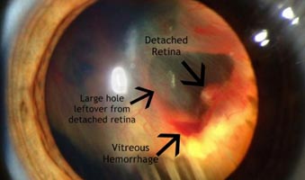
Retinal detachment is an extremely serious eye condition that occurs when the retina gets separated from its underlying tissue within the eye. This makes the retina dysfunctional and can result in permanent vision loss, if proper treatment is not administered yearly.

What are the symptoms?

There is no pain experienced when one is suffering from retinal detachment but warning signs appear much in advance. These include

Photopsia – experiencing sudden but brief flashes of light in the peripheral field of vision. These flashes more commonly occur when the eye moves
retinal detachment Treatment in mumbai
  • The sudden appearance of many floaters — seeing small bits of debris in your field of vision that look like spots, hairs or strings and seem to float before your eyes
  • A shadow or curtain over a portion of your visual field that develops as the detachment progresses
  • Sudden decrease in vision.
  • Straight lines appearing as curved
  • The eye feeling somewhat heavy

If you notice any of the above symptoms, schedule an eye check-up immediately.

What are the causes?

Retinal detachment can occur as a result of

  • Shrinkage or contraction of the vitreous (the gel-like material that fills the inside of your eye) as a part of the normal aging process.
  • Trauma
  • Advanced diabetes
  • High myopia
  • Inflammatory eye disorder

Types of retinal detachment

Rhegmatogenous retinal detachment– a break, tear or hole develops in the retina, allowing liquid to pass from the vitreous space into the sub retinal space between the sensory retina and the retinal pigment epithelium (the pigmented cell layer just outside the neurosensory retina).

Secondary or Exudative or Serous retinal detachment– an inflammation, vascular abnormalities or tumour causes fluid to build up under the retina (there is no hole, break or tear in this case), resulting in retinal detachment.

Tractional retinal detachment– an injury, inflammation or neovascularisation causes the fibro vascular tissue to pull the sensory retina from the retinal pigment epithelium. eg: Diabetic tractional retinal detachment.

Who’s at risk?

You’re more likely to get a detached retina if you

  • Are severely nearsighted (extreme myopia)
  • Have had an eye injury
  • Have a family history of retinal detachment

Note: Retinal tears and retinal detachment are different things. Retinal tears often happen first. If these tears are not treated in time, fluids from within the eye may pass through the tear and separate the retina from its underlying tissue, causing retinal detachment. Retinal detachment may also happen without any warning. But this is more likely in the elderly or those suffering from extreme cases of myopia.

How is retinal detachment diagnosed and treated?

To diagnose retinal detachment, your ophthalmologist would examine the insides of your eyes using an ophthalmoscope. It provides a highly detailed 3-D view, allowing the doctor to see any retinal holes, tears or detachments. Ultrasonography may also be performed which uses sound waves to create the picture of your eye on a video monitor.

To re-attach the retina and prevent future occurrences of retinal detachment, eye surgery needs to be performed. Different modes of treatment include

  • Laser surgery (photocoagulation) – where a laser beam is directed through a contact lens or ophthalmoscope which fuses the tissues back together. This is useful in cases of retinal tears without detachment, or in cases of localized, sub-clinical detachments.
  • Cryopexy (freezing) – also called cryosurgery (cryotherapy), the eye is exposed to extreme cold to destroy the abnormal or diseased tissue. In this case, the procedure produces a delicate scar that helps connect the retina to the wall of the eye.
  • Pneumatic retinopexy – this procedure is more common if the detachment is uncomplicated.
    • The surgeon starts by freezing (cryopexy) the tear area
    • A bubble is injected into the vitreous cavity of the eye, causing the retina to push against the tear and the detached area
    • This continuous pressure (may go on for days) eventually makes the retina re-attach itself to the wall of the back of the eye
  • Scleral buckling – very thin bands of silicone rubber or sponge are sewn onto the sclera (outside white of the eye). The tissue around the area of the tear may be frozen or lasers may be used to scar and re-attachthe tissue.
  • Vitrectomy– the vitreous gel is removed and a gas bubble or silicon oil bubble is used which holds the retina in position. The silicon oil is removed 3 to 6 months after the procedure depending on the condition of the underlying retina.

Tag Cloud