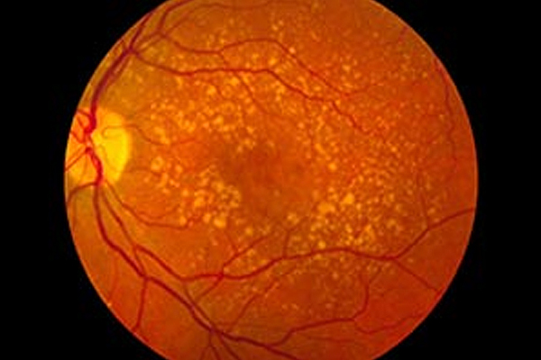
It progressively destroys your sharp central vision. This impairs your ability to see objects clearly and makes it very difficult to undertake the normal tasks such as reading and driving.
Age Related Macular Degeneration affects the macula, the part of the eye that allows you to see things in fine detail. (The macula is located in the centre of the retina, the light-sensitive tissue at the back of the eye.) What you may see
Age Related Macular Degeneration can cause loss of sharp central vision in one or both eyes. With Age Related Macular Degeneration, you may have no obvious vision loss. Or you may have one or more of the following vision problems:
The thought of vision loss can be frightening. You may fear going blind. Or you may worry about being unable to drive, read, or be independent. Although Age Related Macular Degeneration can cause vision loss that ranges from mild to severe, it rarely causes total blindness. What you can do.
Whether you have Age Related Macular Degeneration or are at risk for it, there are ways in which you can protect the vision that you have:
When a loved one has Age Related Macular Degeneration, you can help. Encourage your loved one:
The eye receives and processes light, allowing you to see. Central vision is the sharp detailed vision that you use when you look straight ahead. Peripheral vision is side-vision, the less acute vision that you may call ‘seeing out of the corner of your eye’. Age Related Macular Degeneration affects central vision. Mechanism of sight
The healthy macula The macula is the part of the retina where central vision takes place. The fovea is the most sensitive part of the macula. The whole retina, including the macula and the fovea, has several layers:
There are two kinds of macular degeneration: dry and wet. Age Related Macular Degeneration may be either kind. Dry macular degeneration is more common. It usually does not cause severe vision loss. Wet macular degeneration is less common, but it is more likely to cause severe vision loss. Dry macular degeneration can sometimes progress to wet macular degeneration.
Dry macular degeneration may cause a slow and gradual decline in vision. In this condition, there is formation of drusen (yellow deposits) beneath the retina. These changes may cause distorted or blurry vision. As the function of the macula is impaired over time, the central vision is completely lost in the affected eye.
Amongst these two types of Age Related Macular Degeneration, the dry condition is predominant. Over 85% of all the people who suffer Age Related Macular Degeneration have the dry-type condition. Wet macular degeneration Wet macular degeneration may cause sudden loss of central vision. The growth of new, weak blood vessels causes it. These blood vessels grow from the choroid below through the breaks in the RPE. They may cause the macula to bulge, distorting vision. Fluid leaking from these weak blood vessels, or scarring on the surface of the retina, can cause dark or blurred spots in the field of vision.
Vision monitoring Check your vision often using the Amsler grid. Note changes in your vision or any distortion in your visual field and report them to your eye doctor. Keep in mind that you may have vision problems that are not related to Age Related Macular Degeneration. Making sure that all eye problems are treated adequately, helps you make the most of your remaining vision. The amsler grid An Amsler grid is a chart that you can use at home to check your vision. Cut out the grid below. Or your eye doctor may give you one. Use the grid regularly, as directed by your eye doctor. If you notice vision changes, contact your ophthalmologist as soon as possible.
Your evaluation assesses two things: your vision and the health of your eyes. Vision tests help your doctor learn about how well you see. By examining your eyes your doctor can find out more about your eye health.
Your medical history Your doctor will ask about your symptoms, age and health history. He or she may also ask about the medical history of your family and factors such as smoking, diet, and exercise. He will also undertake a routine eye examination.
Photographs of the retina If your doctor thinks you may have wet Age Related Macular Degeneration, he or she may do an angiogram (a special photograph of the retina assisted by fluorescein dye injection). For this procedure, dye is injected into a vein, usually in the arm or hand. It then travels to the eye. The dye highlights any abnormal blood vessels or leaking fluid.
In this procedure, a laser beam destroys the tiny leaky blood vessels. A high energy beam of laser is directed at the blood vessels to prevent further blood and fluid leakage. This prevents further loss of vision. Only a small percentage of people with wet Age Related Macular Degeneration can be successfully treated with Laser.
Laser surgery is more effective when leaky blood vessels have developed away from the fovea, the central part of the macula. The risk of new blood vessels forming is not eliminated with Laser procedures, and further procedures may be necessary in time to come. Photo-Dynamic Therapy (PDT)
The therapy is painless and can be done within twenty minutes.
Copyright © 1987-2024 Ojas Eye Hospital All rights reserved | Privacy Policy
*Disclaimer: All information on www.lasikindia.com for informational purposes only and is not intended to be a substitute for professional medical advice, diagnosis, or treatment. Always seek the advice of your physician or other qualified health care provider.